The Åstrand Laboratory
At the Åstrand Laboratory, occupational physiology research focuses on physical performance, health, and physical activity. The laboratory is namned after former GIH professor Per-Olof Åstrand.

Research at the laboratory
The research at the Åstrand Laboratory includes:
- the body's adaptation to acute work and training.
- the importance of nutrition for physical performance.
- the muscle's adaptation to endurance training and strength training, physical activity and exercise habits in children and adults, exercise and insulin sensitivity.
- the function of mitochondria in the human musculature.
Physiology and biochemical laboratory
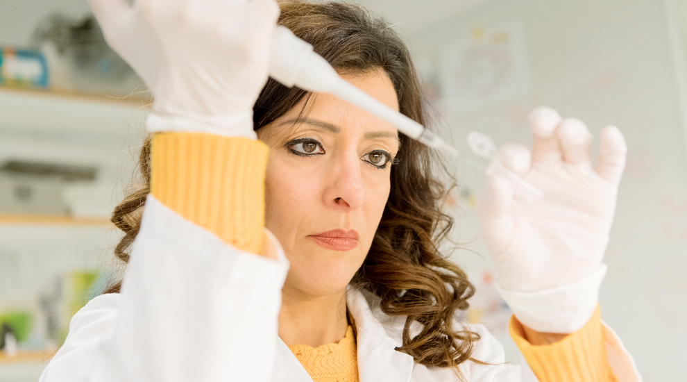
The Åstrand Laboratory is a work physiology and a biochemical laboratory. The work physiology laboratory has equipment for doing work tests, measuring fitness and performance.
The biochemistry laboratory performs histochemical, biochemical and molecular biological analyses on blood and muscle samples. This includes analyzes of muscle fiber composition, enzyme and metabolite determinations, determination of protein expression (Western blot), gene expression (PCR analysis), and measurement of mitochondrial function in isolated muscle preparations.
Tests and methodology at the Åstrand Laboratory
The methodology is used as a traditional determination of oxygen uptake and physical performance, as well as sampling in muscle biopsy and blood tests.
Measurement of maximum oxygen uptake
This test measures your fitness, or more accurately, how much oxygen your body can absorb during maximal work. The test takes about 8–12 minutes and is normally done on a treadmill or ergometer. When running, the load gradually increases from jogging to hard running until you can't take it anymore. During the test, you wear a mask to measure the oxygen content of your exhaled air and the volume of air you breathe in and out per minute (ventilation).
Slightly simplified, your maximum oxygen uptake (in liters per minute) is calculated by multiplying the ventilation by the difference in oxygen content in room air and exhaled air. This value is often divided by the body weight, and the so-called test value is obtained, i.e. maximum oxygen uptake in milliliters per minute and kilogram.
An exerciser is usually between 40-50 ml/min/kg, while well-trained elite athletes in cardio sports have test values of > 70 ml/min/kg. During a maximum test, the heart rate is also recorded continuously.
Calculation of maximum oxygen uptake
If you do not have access to the equipment required to measure maximal oxygen uptake or, for other reasons, are unable to perform a maximal test, there are non-maximal tests. It can be used to calculate the maximum oxygen uptake capacity. Usually, these are done on an ergometer bike.
The Åstrand test is the best-known test for determining fitness. It was developed in the 1960s by husband P-O and and wife Irma Åstrand at GIH.
You can find more information about the Åstrand test in "Åstrand's test handbook".
A further development of the test, the so-called Ekblom-Bak test, was recently presented.
There is a need to reliably measure the daily movement pattern, that is everything from sedentary and low-intensity activity to moderate or high-intensity exercise. Asking a person to report how much they sit or move is simple but unreliable.
At GIH, we have been working for several years to measure the daily activity pattern using objective methods. They are small meters, so-called accelerometers, and inclinometers, which are worn on the hip in a belt, around the wrist, or attached to the thigh.
When a person moves, acceleration occurs, and this is what the accelerometer registers. The more intensely the person moves, the greater the rash. Sitting still or standing still has no effect. If the inclinometer is attached to the thigh, it can determine whether the person is standing or sitting.
In this way, in various studies and situations, we can determine how much a person sits or moves and how the activity or sitting still is distributed over the day or week. We can then describe this or relate it to different outcomes, such as well-being, disease risk, or performance.
In most studies carried out at the Åstrand laboratory, blood samples and muscle samples are taken while they do work trials. Muscle sampling, a so-called biopsy, is a minor surgical procedure. While the skin and muscle surface are numbed, a small incision is made. A needle or forceps is inserted into the muscle, and a small piece is cut out. The small amount of muscle tissue removed does not affect muscle function, and the muscle fibers are repaired after a short period.
The muscle biopsy consists of 200–500 fragments of muscle fibers and weighs between 20 and 80 mg. Typically, muscle biopsies are taken from the outer part of the anterior thigh muscle. In some cases, analyzes are performed on new muscles. Still, the most common is that the muscle biopsy is frozen as quickly as possible in liquid nitrogen and stored at -80 degrees pending analysis.
Measurement of oxygen uptake in vitro
Several studies at the Åstrand laboratory investigated how different exercise types affect the muscle's maximum oxygen consumption. This is done by incubating intact bundles of fibers from a muscle biopsy under standardized conditions, and oxygen consumption is measured after adding various substrates.
Measurements are also carried out on isolated mitochondria. The equipment used is called Oxoboros and provides unique opportunities to investigate how exercise and nutrition affect oxygen uptake in vitro.
The human musculature consists of a mixture of slow (type I) and fast (type II) muscle fibers with different contractile and metabolic properties. Using histochemical methodology, we can obtain information about fiber composition, fiber surfaces, capillary density, and glycogen content in individual fibers.
For this analysis, the muscle biopsy is mounted in a "tissue adhesive" (Tissue Tek), frozen in isopentane, cooled by liquid nitrogen to avoid freeze damage. In a cryostat at -20 °C, thin cross-sections (10 µm) of muscle fibers are cut, fixed on a glass coverslip and then treated differently depending on the subsequent analysis.
When analyzing fiber composition, that is proportion of fast and slow fibres, the knowledge that the enzyme myosin ATPase has different pH sensitivity in other fiber types are used. The enzyme is inactivated in slow fibers at alkaline pH, causing these fibers to appear white, while fast fibers with intact ATPase activity turn black in the final staining procedure.
Analyzes of individual muscle fibers
The histochemical methodology only provides a semi-quantitative measure of substrate content in different fiber types. The muscle biopsy is freeze-dried to quantify glycogen, amino acid concentration, enzyme activity, or gene expression in individual fibers. Individual muscle fibers, which usually weigh only 15 µg, are separated from each other.
Part of the fiber is cut off and identified as type I or type II using ATPase staining; see above. The remaining fiber is used for biochemical or molecular biological analyses performed on single muscle fibers or pools of type-identified fibers.
Glucose, lactate, and hormone levels in plasma are usually analyzed in conjunction with labor trials. The former with enzymatic methodology in a spectrophotometer and hormone levels in a plate reader using ready-made kits.
Muscle glycogen concentration is often analyzed in biopsies taken in connection with exercise to calculate consumption during work. Glycogen is broken down enzymatically in a buffer solution, and the glucose content is then analyzed using the same methodology as glucose in plasma. Measurement of the maximum activity of enzymes that regulate the breakdown of carbohydrates and fat can also be done on human biopsies. The muscle sample is homogenized under optimal conditions and after adding substrate, the reaction rate is followed in a spectrophotometer.
Enzymes with such a low maximum activity that cannot be measured with said methodology are analyzed by labeling the enzyme's substrate with a radioactive isotope, and the conversion to product, after separation from the labeled substrate, is measured for a given time in a scintillation counter.
In most studies at the laboratory, how exercise and nutrition affect the enzyme activity of specific enzymes that regulate various processes in the muscle is investigated. When the activity of an enzyme is low or underlying mechanisms are to be investigated, a methodology called Western blot is used.
The muscle sample is freeze-dried for this analysis and cleared of connective tissue and blood. The sample is then homogenized, and the proteins are separated by SDS-PAGE, a method that separates proteins concerning molecular weight.
Slightly simplified, the proteins migrate at different speeds through a polyacrylamide gel after an electrical voltage is applied. The protein with the lowest molecular weight travels the fastest through the gel.
The next step is to transfer the proteins to a membrane (PVDF) which is then incubated with a specific antibody (primary antibody) against the protein of interest.
Then a secondary antibody is applied that binds to the primary antibody, and the amount of protein is detected after adding a reagent that emits chemiluminescent light.
Exercise increases the gene expression of several proteins that regulate the metabolism of carbohydrates, fat, and protein. Even a single training session can stimulate a particular gene, indicating change after more prolonged training. Stimulation of a specific gene is measured as a change in mRNA expression.
The first step in this analysis is to isolate RNA from the muscle sample, then cDNA is synthesized, which, via cyclic heat treatment, causes the gene to be amplified to measurable mRNA levels. This methodology is called RT-PCR.
Liquid chromatography is used to analyze amino acids in plasma and muscle. It can say that the sample is separated into a column with a specific composition depending on how strongly they bind to the column material.
For the sample to migrate through the column, a so-called mobile phase, usually a solvent, is pumped through the column at a given rate. Different amino acids travel through the column at different speeds and are then detected using a mass spectrometer based on their molecular weight.
The methodology recently developed at the laboratory is called UPLC (Ultra Performance Liquid Chromatography).
On this page
Contact
 Docent, Deputy head of Department, Senior lecturer, Laboratory managerMarcus Mobergmarcus.moberg@gih.se+46 8-120 53 875
The Ekblom-Bak test
The Ekblom-Bak test was developed by researchers at GIH and is a submaximal cycle ergometer test for calculating VO2max.

